Smile Visualization
Take a smiling selfie and we’ll show you what Invisalign® treatment can do for you. Sometimes insurances can cover upto $2500 of invisalign treatment. Call our office or follow the link to find out.
At Fuller Smiles San Fernando Valley, we combine clinical experience with advanced imaging to give patients clearer answers and more predictable treatment outcomes. Cone-beam computed tomography (CBCT) has become a cornerstone of modern dental diagnostics because it produces three-dimensional views of teeth, jaws, and surrounding structures that traditional two-dimensional X-rays cannot show. Our approach is to use CBCT selectively and thoughtfully so each scan provides meaningful information that guides care.
The goal of CBCT is not simply to take more pictures, but to reveal anatomy and relationships that affect diagnosis and treatment planning. When used appropriately, CBCT helps clinicians detect conditions, measure structures precisely, and plan procedures with a higher degree of confidence. Below, we explain what CBCT does, how it supports different types of dental care, and what patients can expect during and after a scan.
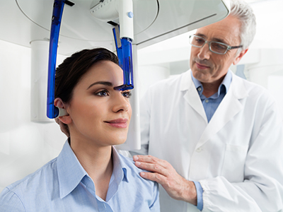
CBCT uses a cone-shaped X-ray beam and a digital detector that rotate around the patient’s head to capture a volumetric dataset. The resulting images can be reconstructed into axial, sagittal, and coronal views as well as 3D renderings, which allow clinicians to examine anatomy from any angle. Unlike traditional panoramic or periapical films, CBCT displays depth, spatial relationships, and the true geometry of complex structures.
The level of detail CBCT provides makes it particularly helpful for identifying small anatomical features such as accessory canals, narrow bone ridges, impacted tooth positions, and the course of nerves. Because clinicians can measure dimensions and angles directly on the dataset, CBCT supports precise decision-making—for example, determining the exact height and width of bone available for an implant or visualizing the position of an unerupted tooth in relation to adjacent roots.
Although the technology produces more comprehensive images than conventional radiography, modern CBCT units are designed to focus on targeted regions and to limit radiation exposure by selecting appropriate fields of view and imaging protocols. This combination of detailed visualization and responsible imaging makes CBCT a powerful diagnostic tool when used for appropriate clinical indications.
CBCT is widely used across many dental specialties because it reveals information that is not visible on flat X-rays. In implant dentistry, CBCT helps assess bone volume, detect critical anatomical landmarks such as the inferior alveolar nerve or maxillary sinus, and plan optimal implant position and angulation. For oral surgery, CBCT clarifies the relationship between impacted teeth and adjacent structures, reducing the risk of complications.
Endodontists and general dentists rely on CBCT to investigate persistent symptoms when conventional films are inconclusive—such as detecting vertical root fractures, assessing complex root canal anatomy, or locating canals that are difficult to visualize. In orthodontics and airway assessment, CBCT contributes to evaluating jaw relationships, tooth impactions, and airway space in three dimensions, supporting more individualized treatment strategies.
CBCT also plays an important role in evaluating pathology and trauma by providing a comprehensive view of fractures, cysts, or lesions that may affect the jaw, teeth, or sinuses. In each of these applications, the value of CBCT lies in its ability to reduce uncertainty, minimize surgical surprises, and improve communication between the clinician and patient.
A CBCT scan is only as useful as the interpretation that follows. Our clinicians pair advanced imaging with clinical examination and patient history to form a complete diagnostic picture. Interpreting volumetric data requires experience and training to recognize normal anatomical variations, artifacts, and clinically relevant findings—skills our team continually refines through ongoing education and case review.
When a scan reveals findings that require specialist input—such as suspicious lesions, complex surgical anatomy, or airway concerns—we coordinate care with oral surgeons, endodontists, or medical professionals as needed. Sharing CBCT datasets with consultants improves treatment coherence and often streamlines next steps because everyone is reviewing the same comprehensive information.
We also use CBCT images as a communication tool during treatment planning. Three-dimensional visuals help patients understand the nature of a problem and the rationale for recommended procedures, which supports informed consent and aligns expectations before any intervention begins.
CBCT imaging is typically quick and noninvasive. A single scan can be completed in under a minute of actual exposure time, with the entire appointment usually lasting only a few minutes for positioning and instructions. Patients remain seated or standing while the scanner rotates, and most find the experience simple and unobtrusive.
Safety is a primary concern with any radiographic procedure. Modern CBCT systems offer adjustable fields of view and exposure settings so clinicians can image only the area of interest at the lowest dose consistent with diagnostic quality. Our team follows accepted radiation-safety principles, including selecting appropriate protocols for children and pregnant patients, and avoids unnecessary imaging.
After the scan, there is no special recovery period—patients can resume normal activities immediately. The clinician will review the images, explain key findings, and discuss how the results inform the proposed treatment plan. If additional evaluation is needed, we will outline the recommended next steps and, when appropriate, arrange coordination with specialists.
When used thoughtfully, CBCT enhances many aspects of dental care by turning uncertainty into actionable information. From implant position guides to surgical planning and complex endodontic diagnosis, three-dimensional imaging supports interventions that are more accurate and tailored to the patient’s anatomy. This precision can reduce procedural time, lower the risk of complications, and improve long-term outcomes.
At the practice level, CBCT integrates with other diagnostic tools—clinical examination, intraoral photography, digital impressions, and conventional radiographs—to provide a complete clinical dataset. Combining these resources allows clinicians to refine treatment sequencing, anticipate anatomic challenges, and create restorations or surgical guides that match the patient’s unique needs.
Fuller Smiles San Fernando Valley is committed to using CBCT responsibly as part of a patient-centered care model. We focus on selecting the right imaging tools for each case, interpreting results with clinical expertise, and communicating findings clearly so patients can make informed decisions about their oral health.
In summary, CBCT is a valuable diagnostic resource that contributes to safer, more precise, and more predictable dental care. If you would like to learn more about how three-dimensional imaging may support your treatment plan, please contact us for more information.

CBCT stands for cone-beam computed tomography, a specialized three-dimensional imaging technology used in dentistry. Unlike standard two-dimensional X-rays, CBCT captures volumetric data that shows teeth, jawbone, airways and surrounding anatomy in true 3D. The scan reconstructs multiple views that clinicians can rotate and measure precisely to support diagnosis and treatment planning.
CBCT produces high-resolution images with focused fields of view tailored to the clinical need, which helps clinicians evaluate complex anatomy more accurately. Because it images structures in three dimensions, CBCT often reveals details that would be difficult or impossible to see on conventional X-rays.
Traditional dental X-rays produce two-dimensional images that compress complex anatomy into a flat view, while CBCT produces a true three-dimensional dataset. This dimensional advantage reduces distortion and overlap, allowing for more accurate assessment of tooth positions, bone volume and nerve pathways. CBCT also allows clinicians to take precise measurements and create cross-sectional views that are invaluable for surgical planning.
Another key difference is the field of view and image reconstruction flexibility; CBCT systems can be adjusted to capture a small region, a quadrant or the entire maxillofacial area depending on the diagnostic need. Although CBCT exposes patients to more radiation than a single periapical X-ray, modern devices and protocols are designed to minimize dose while maximizing diagnostic value.
CBCT is particularly valuable for implant planning, complex extractions, assessment of impacted teeth, evaluation of jaw pathology and treatment of endodontic problems. It provides detailed information about bone quality and quantity, proximity to nerves and sinus anatomy, which improves surgical accuracy and safety. Orthodontic and airway assessments also benefit from 3D imaging when skeletal relationships must be evaluated precisely.
In addition, CBCT can aid in the evaluation of trauma, orthodontic treatment planning and temporomandibular joint (TMJ) assessments by showing bony changes and spatial relationships that are not clear on 2D films. Clinicians use the added information to develop more predictable, individualized treatment plans and to identify issues that might otherwise be missed.
CBCT is considered safe when used appropriately and with justification for the clinical question being asked. Practices follow the principle of ALARA (as low as reasonably achievable) to reduce radiation exposure by using the smallest field of view and lowest effective settings that still deliver diagnostic image quality. Modern CBCT units include low-dose protocols and shielding procedures to protect patients during imaging.
Certain populations, such as pregnant patients or children, require special consideration and CBCT is used only when the diagnostic benefit outweighs any potential risk. Your dentist or imaging specialist will review the need for CBCT and explain how safety measures are applied for your specific case.
A CBCT scan is a quick, noninvasive procedure that typically takes less than a minute for the actual image acquisition. Patients are positioned standing or seated while the scanner rotates around the head; remaining still and following simple breathing instructions helps ensure a clear image. There is no injection or contrast required for routine dental CBCT exams, and most patients experience only minimal discomfort from positioning.
After the scan, the images are processed and reconstructed into 3D views that the clinician reviews to answer the clinical question. The practice staff will explain the next steps and how the images inform your diagnosis and treatment options, so you understand the findings and proposed care plan.
CBCT provides precise measurements of bone height, width and density, and it maps critical anatomical structures such as the inferior alveolar nerve and maxillary sinuses. This detailed information allows clinicians to select the correct implant size, determine optimal angulation and avoid vital structures during surgery. The result is a more predictable surgical plan with fewer surprises at the time of placement.
When indicated, CBCT data can be integrated with digital planning software to create virtual treatment plans and to design surgical guides that translate the plan accurately to the clinical setting. Using this technology leads to more efficient procedures, better primary stability and improved long-term outcomes for implant patients.
Yes, CBCT is effective at identifying many types of bony pathology, including cysts, tumors, inflammatory lesions and fractures. For TMJ evaluation, CBCT shows osseous structures such as condylar shape, joint surface changes and bony asymmetry, which help in diagnosing degenerative or developmental conditions. However, CBCT is limited in soft-tissue contrast, so additional imaging such as MRI may be recommended when soft-tissue or disc evaluation is required.
Because CBCT reveals both localized and broader anatomic changes, clinicians can detect incidental findings that merit further evaluation or referral to a specialist. When abnormalities are identified, the practice communicates findings and coordinates follow-up care or advanced imaging as needed.
CBCT scans are interpreted by trained clinicians who have experience reading volumetric dental images, and complex cases may be reviewed by an oral and maxillofacial radiologist for a formal report. The interpreting clinician evaluates the images in the context of your history and clinical exam to provide clinically relevant findings and recommendations. This interpretive process ensures that the images are translated into a clear diagnosis and an actionable treatment plan.
At Fuller Smiles San Fernando Valley, the treating dentist or specialist will discuss the results with you, explain any recommended next steps and answer questions about diagnosis or treatment options. If a referral or additional imaging is needed, the practice will coordinate that care and provide the necessary documentation to other providers.
CBCT is not a routine screening tool and should be used only when clinically indicated to answer a specific diagnostic or treatment-planning question. The frequency of CBCT exams depends on individual treatment needs, the complexity of care being provided and professional guidelines that prioritize patient safety. Dentists weigh the potential diagnostic benefit against radiation exposure before recommending repeat imaging.
For long-term treatment or monitoring, clinicians document the justification for each CBCT scan in the patient record and consider lower-dose alternatives when appropriate. Patients with evolving conditions will have imaging scheduled based on disease progression, treatment milestones and best-practice protocols.
Clinicians minimize exposure by using the smallest practical field of view, selecting appropriate resolution settings and applying low-dose protocols whenever diagnostic quality permits. The decision to image is always based on clinical justification, and operators follow manufacturer recommendations and regulatory guidelines to ensure safe technique. Proper patient positioning and training of staff also reduce the likelihood of repeat scans due to motion or technical errors.
Additional protections include using lead aprons when appropriate and tailoring imaging protocols for vulnerable populations such as children, who require the lowest effective dose. These measures ensure that CBCT provides maximum diagnostic benefit while adhering to accepted radiation-safety principles.
Take a smiling selfie and we’ll show you what Invisalign® treatment can do for you. Sometimes insurances can cover upto $2500 of invisalign treatment. Call our office or follow the link to find out.