Smile Visualization
Take a smiling selfie and we’ll show you what Invisalign® treatment can do for you. Sometimes insurances can cover upto $2500 of invisalign treatment. Call our office or follow the link to find out.
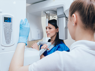
Digital radiography replaces traditional film with electronic sensors and computer processing to capture dental images. Instead of developing X-ray film in a darkroom, a digital sensor records the image and immediately sends it to a computer where it can be displayed, adjusted, and stored. The underlying physics of X-ray production remains the same, but the workflow and image-handling capabilities are dramatically improved. This shift has made radiographic imaging faster, more versatile, and better suited to modern, paperless dental record systems.
The sensor itself is a compact, disposable-friendly device positioned in the mouth much like a film packet. Because images are captured and transmitted electronically, there’s no chemical processing, no physical film to file, and far fewer steps between capture and diagnosis. For dental teams, that means fewer logistical errors and clearer audit trails in the patient record. For patients, it means a more streamlined visit and access to visual explanations right in the treatment room.
Beyond convenience, digital radiography paved the way for further advancements such as three-dimensional imaging and sophisticated image-analysis software. While two-dimensional intraoral sensors remain the workhorse for routine exams, the ability to integrate those images with other digital tools has elevated diagnostic accuracy and treatment predictability across preventive, restorative, and surgical procedures.
One of the most tangible advantages of digital X-rays is improved image quality and manipulation. Clinicians can enhance contrast, zoom in on suspicious areas, and apply filters that make subtle differences more apparent than they might be on film. These capabilities help dentists detect early decay, assess bone levels, evaluate root structures, and identify other pathologies with greater confidence. Enhanced visualization reduces guesswork and supports evidence-based decision making.
Digital images are also easier to compare over time. When successive X-rays are stored in a patient’s electronic record, clinicians can quickly overlay or place images side-by-side to track progression or healing. This is particularly useful for monitoring periodontal health, evaluating the response to endodontic treatment, or following the osseointegration process after implant placement. Consistent image capture protocols make these comparisons reliable and clinically meaningful.
Because images can be shared instantly and securely, collaboration with specialists becomes more efficient. Whether coordinating care with an oral surgeon, periodontist, or orthodontist, digital radiographs facilitate timely consultations and more cohesive treatment planning. The net result is faster, more accurate diagnoses and treatment pathways that are better tailored to each patient’s unique needs.
Patients benefit from digital radiography in several practical ways. Radiation exposure with modern digital sensors is typically lower than with conventional film, largely because sensors are more sensitive and capture usable images with less energy. Dentists also avoid unnecessary repeat exposures by checking image quality instantly; if an image is unsatisfactory, a retake can be done immediately while the patient is still available, rather than requiring a return visit. This efficiency improves both safety and convenience.
Comfort is another frequently noticed improvement. Modern sensors are thinner and ergonomically shaped compared with older film packets, which reduces gagging and discomfort for many patients. Quick image capture shortens the time a sensor needs to remain in the mouth, and the immediate appearance of the image on-screen allows the clinician to walk patients through findings in real time. When patients can see and understand what the clinician sees, they are better able to participate in treatment decisions.
From an environmental perspective, going digital eliminates chemical developers and paper-based processes associated with film. That reduces hazardous waste and the environmental footprint of radiographic services. For patients who appreciate sustainable practice operations, digital radiography represents a meaningful step toward greener healthcare delivery.
At the practice level, digital radiography integrates seamlessly with electronic dental records and imaging software to create a unified clinical workflow. Once captured, images are labeled, filed, and linked to the patient’s chart, making them accessible to any authorized clinician on the team. This structured approach speeds charting, reduces transcription errors, and helps maintain comprehensive records that support continuity of care.
Digital images also serve as powerful educational tools during consultations. Clinicians can highlight areas of concern, annotate images, and show magnified views that help patients understand conditions and proposed treatments. This visual approach often clarifies the rationale for recommended care while building trust through transparency and education. It also supports more precise discussions when coordinating multidisciplinary care or seeking input from outside specialists.
Quality assurance is built into this technology-driven workflow. Sensors are calibrated and cleaned according to infection-control protocols, and staff receive training on positioning and exposure settings to ensure consistent image quality. When combined with routine equipment maintenance and software updates, these practices ensure that imaging remains both reliable and safe for every patient.
Safety and privacy are fundamental to modern dental imaging. Digital radiography equipment is used in accordance with established clinical guidelines to minimize exposure while capturing clinically useful images. Lead aprons and thyroid collars may be employed when appropriate, and exposure settings are adjusted to match the diagnostic need and the patient’s physical characteristics. These measures help ensure that patients receive the imaging necessary for care while keeping radiation exposure as low as reasonably achievable.
On the privacy side, digital images are managed within secure practice systems and handled consistently with healthcare privacy standards. Access controls, user authentication, and secure backup procedures protect patient records and make sure that images are available only to authorized members of the care team or to external clinicians when a patient’s care requires it. These safeguards preserve confidentiality without hindering clinical communication.
Before any imaging, the dental team will explain why specific X-rays are recommended and what the images will help reveal. Patients can expect a brief, comfortable procedure followed by an on-screen review with their clinician. If further imaging is needed for complex cases, the care team will describe the options and how those images will contribute to an effective treatment plan.
Digital radiography has become a cornerstone of modern dental care because it combines precision, safety, and practical benefits for both patients and clinicians. By making images clearer, easier to share, and faster to obtain, this technology supports better diagnoses, more informed treatment planning, and a smoother patient experience. If you’d like to learn more about how digital X-rays are used in our office or whether they’re appropriate for your next visit, please contact us for more information.

Digital radiography uses computer technology and electronic sensors to capture dental images instead of traditional film. These high-resolution images are transferred instantly to a computer where they can be viewed, adjusted, and stored. Because the images are digital, they can be enhanced to improve diagnostic visibility without repeating the exposure.
This method streamlines recordkeeping and enables immediate comparison with prior images for tracking changes over time. It also supports efficient communication between dental professionals because files can be shared electronically when referrals or consultations are needed. Overall, digital radiography is a central diagnostic tool in modern dentistry.
The primary difference is the capture medium: digital sensors replace light-sensitive film, eliminating chemical processing. Digital systems typically require a fraction of the radiation used for conventional film x-rays, while producing images that are available instantly. Clinicians can adjust contrast, magnify regions of interest, and take measurements on-screen, which enhances diagnostic accuracy.
Digital files are easier to archive and retrieve, reducing the risk of lost or damaged films. Because multiple clinicians can view the same digital image simultaneously, coordination of care becomes faster and more convenient. The lack of film processing chemicals also reduces environmental impact in the office.
Digital radiography significantly reduces radiation exposure compared with older film technologies because sensors are more sensitive. Modern digital units and shielding protocols further lower dose while preserving diagnostic image quality. Dentists follow the ALARA principle—keeping exposures as low as reasonably achievable—tailoring imaging to each patient's needs.
For pregnant patients or those with specific medical concerns, clinicians will evaluate risk and may defer or modify imaging when appropriate. Lead aprons and thyroid collars are available for added protection during exams. Open communication with the dental team helps ensure imaging is performed only when clinically necessary.
Digital radiographs reveal problems that are not visible during a visual exam, such as interproximal cavities, bone loss from periodontal disease, and hidden infections at tooth roots. They also identify impacted teeth, evaluate the position of unerupted teeth, and check the integrity of existing restorations. Endodontic issues like periapical lesions can be detected early, supporting timely treatment decisions.
Three-dimensional CBCT imaging, a form of digital radiography, provides cross-sectional views useful for implant planning and complex surgical assessments. While two-dimensional digital x-rays remain the standard for routine screening, CBCT is reserved for cases where detailed spatial information is required. Your dentist will recommend the right type of imaging based on the clinical question and patient history.
Digital radiography is integral to diagnosis and treatment planning because it lets clinicians visualize the anatomy and monitor changes over time. Images inform decisions for restorations, root canal therapy, periodontal treatment, and surgical procedures. Dentists can measure bone levels, assess tooth structure, and verify fit and placement of restorations with on-screen tools.
At Fuller Smiles San Fernando Valley, clinicians combine digital images with clinical findings to develop predictable, evidence-based treatment plans. Digital records streamline follow-up care, enabling comparisons between pre-treatment and post-treatment images. This visual documentation helps clinicians explain options and expected results to patients in clear, evidence-based terms.
A typical intraoral digital radiograph takes only seconds to capture and appears on the computer screen immediately afterward. Patients remain seated while the sensor is briefly positioned in the mouth, and the process is noninvasive and quick. If multiple views are needed, the entire imaging portion of an exam is usually complete within a few minutes.
Images may be enlarged or annotated by the dentist to clarify findings during discussion, and patients can view the same images on-screen. This immediate feedback helps patients understand diagnosed conditions and recommended treatments. If additional imaging such as CBCT is needed, the team will explain what to expect before proceeding.
Digital radiographs are easily shared with other dental offices, specialists, or laboratories using secure electronic transfers or by providing image files on request. This capability expedites referrals, second opinions, and collaborative treatment planning. Sharing also reduces the need for repeat exposures when diagnostic images are already available.
Before transferring images, the practice confirms that electronic exchanges meet privacy and security standards. When patients change providers, copies of their digital records can be requested to ensure continuity of care. Clear communication about what will be shared helps maintain trust between patients and clinicians.
Digital images are stored in electronic health records and protected by the same privacy safeguards that apply to other medical information. Access is restricted to authorized personnel, and systems use secure logins and controlled permissions to prevent unauthorized viewing. Regular backups and IT safeguards help prevent data loss while maintaining the integrity of patient records.
When images are shared with outside providers, the practice uses secure transfer methods and follows applicable regulations to protect patient confidentiality. Patients may ask the office how their images are stored and who can access them if they have concerns. Transparent policies and staff training are key parts of maintaining strong data protection practices.
The need for radiographic imaging is determined by clinical examination, patient history, and risk factors rather than a one-size-fits-all schedule. For patients with active dental problems or higher risk of decay or periodontal disease, more frequent imaging may be warranted, while low-risk patients may require x-rays less often. Dentists balance the diagnostic benefits with radiation exposure to choose the appropriate timing and type of imaging.
Children, adults with missing teeth, and those with significant dental work are evaluated individually to ensure images provide the information needed for safe, effective care. If you have questions about why an image is recommended, ask your clinician to explain the anticipated diagnostic value and any alternatives. Informed consent and shared decision-making help patients feel comfortable with imaging decisions.
Fuller Smiles San Fernando Valley uses modern digital radiography systems in its offices to support accurate diagnosis and efficient recordkeeping. Our clinicians integrate digital imaging into routine exams, restorative care, and advanced procedures when imaging is needed to guide treatment. Using digital tools helps the team communicate findings clearly with patients and coordinate care across specialists when needed.
If you would like to learn more about imaging options or review recent radiographs, the team can explain how images were taken and what they show. Appointments at our Northridge and West Hills locations include access to digital imaging when clinically appropriate. Contact the office to discuss any special imaging needs or to request copies of your digital records.
Take a smiling selfie and we’ll show you what Invisalign® treatment can do for you. Sometimes insurances can cover upto $2500 of invisalign treatment. Call our office or follow the link to find out.