Smile Visualization
Take a smiling selfie and we’ll show you what Invisalign® treatment can do for you. Sometimes insurances can cover upto $2500 of invisalign treatment. Call our office or follow the link to find out.
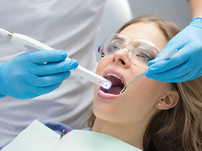
An intraoral camera is a compact, pen-sized device designed to capture high-resolution, full-color images from inside the mouth. Unlike external cameras or X-ray machines, this tool provides a live visual of tooth surfaces, gum tissue, and other oral structures at a magnification level that is easy for both clinicians and patients to see. Its small size and maneuverability make it possible to photograph areas that are otherwise difficult to view during a standard exam.
These images are displayed in real time on a monitor, giving patients an immediate, clear perspective of what the dentist observes. For many people, seeing a crisp visual is far more informative than hearing a verbal description alone. The intraoral camera bridges the communication gap between clinical findings and patient understanding, helping the dental team explain conditions clearly and collaboratively plan next steps.
Although the technology is sophisticated, the experience for the patient is straightforward and noninvasive. The camera is gently guided around the mouth while the clinician captures still photos or short video clips. Those images are then stored in the patient’s digital chart as part of their permanent record, supporting continuity of care and future comparisons.
High-resolution intraoral images allow clinicians to spot early signs of decay, hairline fractures, worn restorations, and soft-tissue changes that might be missed during a visual exam alone. The magnified, well-lit views make subtle defects more apparent, which can lead to more accurate diagnoses and earlier intervention when appropriate. This level of detail improves clinical decision-making without increasing patient discomfort or exposure to radiation.
The ability to zoom in on particular areas and capture multiple angles helps the dental team form a more complete picture of oral health. Intraoral imaging complements traditional diagnostic tools such as radiographs and clinical probing, providing surface-level context that radiographs alone cannot. Together, these modalities create a fuller, more reliable assessment of a patient’s needs.
For patients who experience anxiety or doubt, seeing clear visual evidence can make the diagnosis feel more tangible and less abstract. When the problem is shown rather than simply described, patients often have an easier time weighing treatment options and consenting to recommended care.
One of the most immediate benefits of intraoral cameras is improved patient education. Visual aids help clinicians explain conditions like enamel erosion, gum inflammation, or failing dental work in concrete terms. When patients can view their own teeth on a screen, discussions about oral hygiene, preventive measures, and restorative choices become more meaningful and easier to follow.
Images captured during a visit can be annotated, compared side-by-side with previous photos, or paused to highlight areas of concern. This visual record supports ongoing conversations about progress and goals, whether the focus is preventive care, restorative treatment, or cosmetic improvements. Clear images also make it simpler to demonstrate the reasons behind specific recommendations.
Because these visuals are stored digitally, patients can take a more active role in their care. They leave appointments with a clearer understanding of their condition and the rationale for any suggested procedures, which tends to increase trust and long-term adherence to preventive habits.
Intraoral camera images become an integral part of the clinical record, offering a precise snapshot of oral conditions at a given time. These snapshots are useful for tracking changes, documenting baseline health, and monitoring the outcome of treatments. Having clear photographic documentation supports continuity of care and makes follow-up appointments more efficient and focused.
Captured images are also valuable when collaborating with dental specialists, laboratories, or other members of the care team. High-quality photos can be shared with colleagues to aid in referrals, lab prescriptions, or multidisciplinary treatment planning. This shared visual language reduces ambiguity and helps ensure that everyone involved has the same clear understanding of the clinical situation.
For administrative and regulatory purposes, documented images can clarify the clinical rationale for certain procedures or referrals. Because the photos are securely stored within the patient’s chart, they can be retrieved and reviewed whenever needed to support clinical notes and long-term care strategies.
The intraoral camera is a safe, painless tool with no exposure to ionizing radiation. It is used much like a small mirror but with the added advantage of magnified, high-definition display. Clinicians handle the device with care and maintain strict infection-control protocols, including barrier protections and thorough sterilization of reusable components per accepted clinical guidelines.
During a typical appointment, the dentist or hygienist will move the camera gently along the teeth and gumline to capture a series of images. The process usually takes only a few minutes and can be performed alongside a routine cleaning or exam. Patients who have limited mouth opening or sensitive gag reflexes should mention this to the clinician so the team can adapt technique and ensure comfort.
Because the images are recorded instantly, they can be reviewed with the patient during the visit. This immediate feedback helps patients understand the condition of their oral tissues and gives clinicians the chance to explain targeted steps for treatment or prevention in accessible language.
An intraoral camera is a practical, patient-friendly tool that enhances diagnosis, improves communication, and strengthens clinical documentation. By making oral conditions visible and understandable, this technology supports more informed decision-making and clearer coordination among providers. The practice’s use of intraoral imaging reflects a commitment to modern, transparent care that centers on patient understanding and comfort.
If you’d like to learn more about how intraoral cameras are used during exams or how the technology might benefit your care, please contact us for more information.

Intraoral camera technology refers to a small, pen-sized camera designed to capture high-resolution images inside the mouth and display them on a computer screen in real time. The device provides a close-up, full-color view of teeth, gums and other soft tissues to support clinical examination. Because the images appear instantly on a monitor, patients and clinicians can review oral findings together during the appointment.
The camera can record still images and short video clips that become part of the patient record, improving documentation and continuity of care. These images help clinicians track changes over time and create a visual baseline for comparison at follow-up visits. High-quality intraoral images also support accurate communication with dental laboratories and specialist colleagues when coordinated care is required.
An intraoral camera gives patients a clear visual of clinical findings that are otherwise hard to see, which makes explanations more concrete and easier to understand. Seeing an enlarged image of a cracked tooth, stain or early decay helps you participate in decisions about diagnosis and next steps. This direct view also reduces uncertainty and allows the clinician to point out specific areas of concern.
At Fuller Smiles San Fernando Valley clinicians routinely use intraoral images to walk patients through their oral health status and proposed treatment options. Images can be annotated or saved to illustrate progress, which reinforces education and informed consent. The visual record supports collaborative treatment planning and improves overall patient engagement.
Intraoral cameras capture still photos or short videos that are routed directly into the dental practice management or imaging software during the exam. The clinician positions the device to image a single tooth, quadrant or soft tissue area and captures multiple views as needed to document the condition. Image settings and file formats are optimized for clinical use so files remain clear and useful for diagnosis.
Captured images are saved to the patient’s digital chart as part of their permanent record and are retrievable for future appointments. When indicated, those images can be exported to a secure format for referral, laboratory prescriptions or consultation with specialists. The digital workflow reduces paper records and streamlines interdisciplinary communication while preserving clinical detail.
Yes; intraoral cameras magnify the oral structures and reveal subtle changes in enamel, soft tissue texture and restorative margins that are difficult to see with the naked eye. They are especially useful for identifying surface discoloration, hairline cracks, worn restorations and early signs of decay on accessible surfaces. While the camera improves visual detection, it is used alongside tactile examination and diagnostic radiographs to confirm findings.
By documenting small changes over successive visits, intraoral images allow clinicians to monitor progression and intervene earlier when appropriate. Early detection supports conservative treatments that preserve natural tooth structure and reduce the need for more extensive procedures later on. The camera is therefore an important component of a comprehensive preventive and diagnostic strategy.
An intraoral camera complements radiographs, cone beam CT, digital impressions and other imaging modalities by providing detailed surface views in real time. While X-rays show internal anatomy and bone relationships, the intraoral camera documents external tooth surfaces, soft tissue conditions and restoration margins. Combining surface photography with three-dimensional and radiographic data gives the clinician a fuller picture for diagnosis and planning.
In restorative and prosthetic cases, intraoral images can be sent to the dental laboratory with digital impressions and prescriptions to ensure accurate shade matching and fit. For implant and surgical planning, photographic documentation helps verify soft tissue contours and healing at follow-up appointments. The integrated approach improves coordination, reduces remakes and enhances clinical predictability.
Yes, using an intraoral camera is a noninvasive and generally comfortable procedure for patients of all ages. The device is small and lightweight, and the clinician maneuvers it gently to capture images without causing pain. Most patients experience only minor sensations from positioning and brief retraction of cheek or tongue when necessary.
Clinics follow strict infection-control protocols for intraoral cameras, including barrier sleeves and routine cleaning to meet sterilization standards. Devices are handled by trained staff who adhere to best practices for sterilization and cross-contamination prevention. Because the camera does not emit ionizing radiation, it is safe to use repeatedly as part of routine exams and documentation.
Dental images from an intraoral camera are routinely shared with specialists, dental laboratories and other providers when it is necessary for diagnosis, treatment coordination or laboratory work. Sharing typically occurs with your consent and is limited to the information needed to facilitate care. Images provide a clear visual reference that helps other professionals understand specific clinical issues when preparing treatment or restorations.
The practice uses secure methods to transfer images and accompanying clinical notes so that only authorized recipients receive the information. When images are requested for external review, staff follow established protocols to document the transfer and ensure appropriate use. If you have questions about who will receive your images, ask your clinician and they will explain the process and obtain any necessary permissions.
Intraoral images provide a precise visual record that clinicians use to evaluate current conditions, document treatment needs and illustrate proposed procedures. High-resolution photographs reveal relationships between teeth and soft tissue and help identify margins, contacts and areas requiring restoration or maintenance. These visuals make it easier to discuss alternatives and select the most appropriate clinical approach for each patient.
For complex or multi-step treatments, images serve as checkpoints to confirm progress and refine the plan as needed. They are also valuable when coordinating care among specialists, because all parties can review the same visual evidence. The net result is more predictable treatment outcomes and clearer communication throughout the care process.
Capturing intraoral camera images usually adds only a few minutes to a standard dental exam, depending on the number of views needed and the complexity of the case. For a routine screening the clinician may take a handful of targeted images, which can be completed quickly and incorporated into the chart. When detailed documentation is required for treatment planning or laboratory work, additional images may be captured to ensure comprehensive coverage.
Because the process is streamlined within a digital workflow, image capture, annotation and storage are efficient and do not significantly extend chair time. Patients can review images with the clinician immediately, which often speeds decision-making and informed consent. If you have concerns about appointment length, the team will outline the steps and expected time before proceeding.
Fuller Smiles San Fernando Valley maintains secure digital systems to protect patient records, including intraoral images, using access controls and industry-standard security measures. Electronic patient records are stored on encrypted systems with role-based access so only authorized team members can view or modify clinical files. Regular staff training reinforces confidentiality protocols and ensures that privacy practices align with applicable regulations.
When images must be shared for referrals or laboratory work the practice follows documented procedures to limit disclosure to the minimum necessary information and to obtain patient consent when required. Patients may request copies of their images or ask questions about data use and retention, and staff are available to explain how records are managed. These safeguards help preserve patient privacy while enabling high-quality, coordinated dental care.
Take a smiling selfie and we’ll show you what Invisalign® treatment can do for you. Sometimes insurances can cover upto $2500 of invisalign treatment. Call our office or follow the link to find out.