Smile Visualization
Take a smiling selfie and we’ll show you what Invisalign® treatment can do for you. Sometimes insurances can cover upto $2500 of invisalign treatment. Call our office or follow the link to find out.
Oral cancer refers to malignant growths that develop in the tissues of the mouth and throat. While it represents a small percentage of all cancers diagnosed each year, its location can make it particularly serious because tumors discovered at an advanced stage are harder to treat. Early detection directly improves the range of treatment options and the likelihood of a positive outcome, which is why screening is an essential component of routine dental care.
In the United States, health organizations estimate that roughly 50,000 people will be diagnosed with cancers of the oral cavity and oropharynx annually, and several thousand lives are lost each year to the disease. Although overall mortality has fallen somewhat thanks to improved detection and treatments, the incidence of oropharyngeal cancers linked to the human papillomavirus (HPV) has been rising, changing the landscape of risk in recent years.
Oral cancer can appear in many locations inside the mouth and throat. Common sites include the sides and base of the tongue, the tonsillar region and oropharynx, the gums, the floor of the mouth, lips, inner cheek lining and the hard palate. Because lesions can be subtle in early stages, a careful, methodical screening process during routine exams is the best way to catch suspicious changes before they progress.
For patients, understanding what oral cancer looks and feels like—and why regular screening matters—helps turn passive visits into proactive health care. Dental professionals play a key role not only in examining oral tissues but also in educating patients about signs, risk factors and next steps when abnormalities are found.
Certain behaviors and exposures increase the likelihood of developing oral cancer. Tobacco use, including cigarettes, cigars, pipes and smokeless tobacco, remains one of the most significant risk factors. Heavy alcohol consumption compounds that risk; together, smoking and drinking produce a much higher likelihood of malignancy than either factor alone. Because these exposures are common, screening focuses heavily on identifying changes in people who use tobacco or alcohol regularly.
Age and sex also influence risk. Historically, men—particularly those over age 50—have experienced higher rates of oral cancer than women, although this gap has narrowed in some populations. Another major shift in recent decades is the role of HPV, especially HPV-16, which has been linked to an increase in oropharyngeal cancers. This form of the disease often occurs in younger, otherwise healthy adults and is not directly related to traditional lifestyle risks.
Beyond tobacco, alcohol and HPV, other contributors include chronic sun exposure (which can affect the lips), past radiation to the head and neck, chronic gastroesophageal reflux disease (GERD), certain workplace chemical exposures, and nutritional deficiencies. Some factors, such as genetics and past medical history, aren’t modifiable, but recognizing them allows clinicians to tailor monitoring and follow-up.
Understanding which risk factors apply to you helps your dental team prioritize which areas to inspect most closely and how frequently to reassess. Risk assessment is an important first step in a practical, patient-centered screening program.
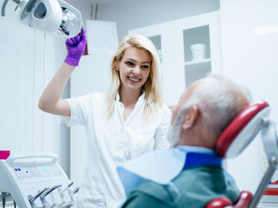
An oral cancer screening is a systematic evaluation performed during a routine exam and typically takes only a few minutes. Your clinician will begin by reviewing your medical and dental history and asking about recent changes in your health or symptoms you’ve noticed. This conversation helps the practitioner identify potential red flags and focus the physical exam more effectively.
The physical portion of the screening includes both visual inspection and manual palpation. The dentist or hygienist examines the lips, tongue (top, sides and under the tongue), gums, inner cheeks, floor and roof of the mouth, and the back of the throat. They will also feel the jaw and neck for any lumps or asymmetry—swollen lymph nodes, for example, can be an early indicator that further evaluation is needed.
Many practices also use adjunctive tools to supplement the standard exam. Specialized lights and tissue visualization devices can make subtle lesions more apparent, and technologies like VELscope® are available in some offices to highlight tissue abnormalities that may not be obvious under normal lighting. These tools do not replace a careful clinical exam but can improve the clinician’s ability to detect suspicious changes.
If a suspicious area is found, the usual next steps are documentation, close monitoring, and referral. Documentation may include photographs or notes about size, color and texture. Depending on the findings, your dentist may recommend a recheck after a short interval or refer you to an oral surgeon, ENT specialist or another physician for further diagnostic tests, such as a biopsy, which is the definitive method to diagnose cancerous changes.
Many early warning signs of oral cancer are subtle or may resemble noncancerous conditions, which is why any persistent or unusual oral change warrants attention. Common signs include sores that do not heal within two weeks, patches of red or white tissue, lumps or thickened areas, and any change in the texture of oral mucosa. Persistent pain or numbness in the mouth or face can also be a symptom.
Other symptoms that should prompt an evaluation are difficulty chewing or swallowing, a feeling that something is caught in the throat, unexplained bleeding, loose teeth not attributable to dental disease, changes in voice, or ear pain without an obvious ear issue. Because these signs can overlap with routine dental problems, context, duration and progression are important factors in deciding whether additional assessment is needed.
Self-monitoring is a useful complement to professional screening. Visitors can perform a brief monthly check by looking inside the mouth with good lighting and feeling around the lips, gums and tongue for any new lumps or patches. If you notice anything persistent or changing, bring it to your dentist’s attention—early evaluation helps separate benign conditions from those requiring urgent care.
Oral cancer screening is most effective when it is part of an integrated, ongoing dental care plan. Because early changes can be hard to detect, consistent follow-up—guided by your clinician’s assessment of your risk profile—ensures that suspicious findings are tracked and managed appropriately. Regular exams give clinicians a reliable baseline, making it easier to spot subtler changes over time.
When screening identifies a concern, prompt referral and coordinated care with medical specialists are critical. Dental teams work with oral surgeons, otolaryngologists and oncologists to confirm diagnoses and plan treatment, and they continue to support patients through recovery, rehabilitation and surveillance afterward. This multidisciplinary approach helps patients navigate complex care pathways with clarity and continuity.
At Fuller Smiles San Fernando Valley, our clinicians incorporate oral cancer checks into routine exams and use available adjuncts to improve detection when indicated. We emphasize clear communication about findings and next steps so patients can make informed choices about follow-up testing and treatment. Prioritizing screening as part of comprehensive preventive care increases the chances of catching disease early when outcomes are best.
In summary, oral cancer screening is a simple yet vital part of routine dental care. By combining a careful clinical exam, awareness of risk factors and timely follow-up when abnormalities appear, dental teams help protect patients and preserve long-term oral health. If you have questions about screening or want to learn more about our approach, please contact us for more information.

An oral cancer screening is a focused clinical exam designed to detect early signs of cancer or pre-cancerous changes in the mouth and throat. The goal is to identify abnormalities such as persistent sores, unusual patches, or lumps before they progress, improving the chances for less invasive treatment. Screenings are preventive measures that complement routine dental care and patient education.
Screening does not diagnose cancer by itself but identifies suspicious findings that need further evaluation. When something unusual is found, your dental team will explain the next steps, which may include close monitoring, imaging or referral for a biopsy. Early identification allows the dental and medical teams to coordinate care promptly and effectively.
Certain factors increase the likelihood of developing oral cancer, including tobacco use, heavy alcohol consumption and a history of significant sun exposure to the lips. Men are more likely than women to be diagnosed, and risk generally rises with age, especially after age 50. Previous head and neck radiation, exposure to certain industrial chemicals, chronic reflux and poor nutrition can also increase risk.
Human papillomavirus (HPV), particularly HPV‑16, has become an important risk factor for oropharyngeal cancers and is often associated with younger patients and those without traditional tobacco histories. Because risk varies between individuals, your dentist will consider your personal and family medical history when recommending the frequency and type of screening.
Seek prompt evaluation for any sore, lump or lesion in the mouth that does not heal within two weeks, especially if it is painless or recurrent. Other warning signs include persistent red or white patches, unexplained bleeding, numbness, a lump in the neck, difficulty swallowing and changes in speech or voice. Any new, persistent oral pain or a loose tooth without an obvious cause should also be checked.
Because early lesions can be subtle, it is important to report even minor or intermittent changes to your dental provider. Timely assessment helps distinguish benign conditions from those that require further testing and ensures appropriate follow-up or referral when needed.
A typical screening begins with a review of your medical and dental history and questions about symptoms or risk factors. The clinician then performs a systematic visual inspection of the lips, cheeks, tongue, floor and roof of the mouth, and examines the throat and oropharynx when feasible. Palpation of the floor of mouth, tongue and neck checks for lumps, firmness or enlarged lymph nodes.
Screenings are noninvasive and usually completed within a few minutes as part of a routine dental exam, although the exact steps may vary depending on your risk profile. If the clinician identifies anything suspicious, they will explain recommended next steps, which may include short‑term surveillance, diagnostic imaging or referral for biopsy.
Most dental professionals include an oral cancer screening as part of your routine dental exam, which commonly occurs every six to twelve months. For many patients, an annual screening is the baseline, while those with higher risk factors — such as tobacco or heavy alcohol use, a history of head and neck radiation, or prior oral lesions — may be screened more frequently. Your dental provider will tailor the screening schedule based on your medical history and current findings.
If you notice any new or persistent oral symptoms between regular visits, you should contact the dental office for an earlier evaluation rather than waiting for your next scheduled exam. Prompt assessment of changes improves the likelihood of early detection and more effective treatment if needed.
Yes — many oral cancers and pre-cancerous lesions can be detected at an early stage through routine visual and tactile screening. Early detection often allows for less extensive treatment, a higher likelihood of successful outcomes and a lower risk of spread to nearby tissues or lymph nodes. Detecting disease early also increases the range of therapeutic options and may reduce the impact on speech, swallowing and appearance.
Because early changes can be subtle, regular dental visits and attention to symptoms are key to timely recognition. When a suspicious finding is identified, prompt diagnostic follow-up, including biopsy if indicated, helps determine the appropriate treatment pathway and improves coordination between dental and medical specialists.
HPV vaccination reduces the risk of infection with the highest‑risk HPV types and is an important preventive measure recommended by public health authorities. If you have questions about HPV, vaccination or its relationship to oropharyngeal cancer, discuss them with your dental or medical provider so you can make informed decisions about prevention and screening.
In addition to visual inspection and palpation, dentists may use adjunctive tools that help highlight abnormal tissue, such as tissue‑fluorescence devices or intraoral cameras. One example is the VELscope® system, which can make abnormal mucosa appear differently under specific light wavelengths; such technologies are intended to supplement, not replace, the clinical exam. These aids can improve visualization and documentation of suspicious areas for monitoring or referral.
When adjunctive findings are unclear, the clinician may recommend further testing, photographic records or referral for diagnostic biopsy. Technology is most effective when used alongside a thorough history and hands‑on exam performed by an experienced clinician.
If your dentist identifies an abnormal finding, they will explain the nature of the finding, the level of concern and recommended next steps. Options may include short‑interval re‑evaluation to see if the lesion resolves, referral to a specialist for biopsy or imaging, or coordination of care with your primary medical provider. Clear communication and documentation help ensure that the issue is tracked and addressed without delay.
It is important to follow the recommended plan and attend any follow‑up appointments so that suspicious lesions are monitored or diagnosed promptly. Early action improves the range of treatment options and the likelihood of a favorable outcome if a serious condition is present.
At Fuller Smiles San Fernando Valley we include a careful oral cancer screening as part of every comprehensive exam, taking a full medical and dental history and performing a systematic visual and tactile evaluation. Our clinicians use clinical judgement to determine when adjunctive devices such as VELscope are appropriate and document any findings for follow‑up. We emphasize patient education so individuals understand risk factors, warning signs and the importance of routine surveillance.
When an abnormality requires further assessment, our team coordinates timely referrals to oral surgeons or ENT specialists and communicates clearly with patients about next steps. Patients seen at our Northridge and West Hills offices can expect a consistent, safety‑focused approach to screening and follow‑up care that prioritizes early detection and coordinated treatment when necessary.
Take a smiling selfie and we’ll show you what Invisalign® treatment can do for you. Sometimes insurances can cover upto $2500 of invisalign treatment. Call our office or follow the link to find out.