Smile Visualization
Take a smiling selfie and we’ll show you what Invisalign® treatment can do for you. Sometimes insurances can cover upto $2500 of invisalign treatment. Call our office or follow the link to find out.
Oral and oropharyngeal cancers are among the cancers for which early diagnosis most directly improves outcomes. When precancerous changes or small lesions are identified before they progress, treatment options are broader and less invasive, recovery is typically quicker, and the chances of long-term survival are substantially better. For patients, this means that routine screening is not simply a formality — it can be a decisive step in preserving health and quality of life.
Regular dental visits create repeated opportunities to spot subtle changes in the mouth that a patient might not notice at home. Because many early abnormalities are painless or visually inconspicuous, relying only on self-examination can leave concerning tissue changes undetected for months. Incorporating advanced screening technology into standard exams helps close that gap and supports proactive care.
Importantly, screening is a preventive habit rather than a diagnostic endpoint. Detecting an irregularity is the beginning of a clinical pathway: additional evaluation, careful monitoring, or referral for biopsy as needed. That pathway is designed to balance timely intervention with avoidance of unnecessary procedures, always prioritizing patient safety and clear communication.
The VELscope® system is an adjunctive device that highlights differences in tissue fluorescence under a safe blue light. Healthy oral mucosa emits a characteristic autofluorescence pattern when illuminated, while many abnormal tissues reflect that light differently. By making those contrasts visible at chairside, VELscope® helps clinicians locate areas that merit closer inspection beyond the naked-eye exam.
It’s important to understand that VELscope® does not provide a diagnosis on its own. Instead, it acts as a visual aid that can reveal suspicious patches, discolorations, or textural changes that could otherwise be missed during a standard inspection. When the device flags an area, the clinician uses clinical judgment — combining the light findings with palpation, medical history, and other observations — to determine next steps.
For patients, the advantage is a more thorough, evidence-informed screening process during routine checkups. The VELscope® exam is noninvasive, quick, and typically added seamlessly into comprehensive oral health visits so that patients receive an elevated level of vigilance without significant extra time or discomfort.
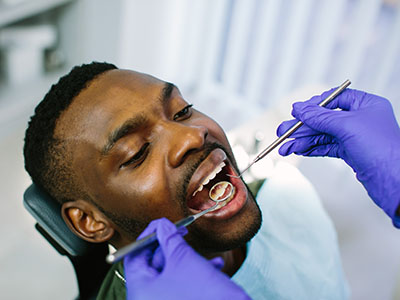
A VELscope® screening begins with a conventional oral exam. Your dental clinician will visually inspect the lips, tongue, floor of mouth, cheeks, palate, and gums, and palpate areas where tissue changes are of concern. Once the preliminary exam is complete, the room light is dimmed and the handheld VELscope® unit is used to examine the same tissues under blue light.
During the light inspection you’ll simply be asked to open wide and move your tongue as directed; there is no contact, no discomfort, and the whole process usually takes only a few minutes. Clinicians look for areas where fluorescence appears diminished, absent, or altered compared with normal tissue. These visual cues guide a careful reassessment of any questionable regions.
If an area appears unusual under VELscope® but looks normal otherwise, your provider may choose to monitor it at follow-up visits. If the finding is suspicious based on combined clinical evidence, the next steps can include targeted imaging, referral to an oral medicine or ENT specialist, or biopsy to obtain a definitive diagnosis. Throughout, clinicians aim to explain observations clearly and discuss appropriate, individualized options.
VELscope® screening can be beneficial for a broad cross-section of patients, but it is especially helpful for those with elevated risk factors. Tobacco and heavy alcohol use, a history of head and neck cancer, significant sun exposure to the lips, or chronic irritation in the mouth can increase risk. Additionally, patients with a persistent sore, lump, or a lesion that does not heal within two weeks should be evaluated promptly.
That said, the device is also useful in routine preventive care because many oral cancers arise without prominent symptoms in the earliest stages. Incorporating VELscope® into regular hygiene visits enables clinicians to build a record of tissue health over time, helping to distinguish transient changes from persistent concerns.
Age and medical history matter as well: adults over 40 and those with immunosuppression or HPV-related risk factors may benefit from closer surveillance. Your dental team will recommend a screening cadence tailored to your personal risk profile and oral health history, ensuring that any monitoring is proportionate and evidence-based.
In our office, VELscope® is one element of a layered approach to oral cancer screening. We combine a detailed clinical exam, patient education, and risk assessment to create a clear plan for monitoring or referral when needed. The goal is to be vigilant without creating unnecessary alarm — emphasizing clarity, timely action, and compassionate communication at every step.
If a suspicious area is identified, clinicians will explain the finding, document it with photos when appropriate, and outline recommended next steps. Those steps may include short-term re-evaluation, referral to a specialist for further assessment, or coordination with a medical team for biopsy and pathology. Throughout the process, our focus is on ensuring patients understand the rationale behind each recommendation.
Fuller Smiles San Fernando Valley integrates VELscope® screening into routine care to give patients the benefit of more sensitive visual assessment during their visits. This technology enhances the diagnostic toolkit of the clinician but does not replace clinical expertise; it supports informed decision-making and timely follow-up when tissue changes warrant further investigation.
In summary, VELscope® Cancer Screening is a fast, noninvasive adjunct that strengthens traditional oral exams by revealing subtle fluorescence changes that merit attention. When combined with consistent checkups, patient education, and appropriate follow-up, it helps create a proactive framework for early detection and better outcomes. If you have questions about how a VELscope® screening is performed or whether it’s appropriate for you, please contact us for more information.

VELscope® oral cancer screening is a chairside examination that uses a specialized blue light to help clinicians visualize abnormalities in the oral mucosa. The device enhances natural tissue fluorescence so that healthy and suspicious tissues appear differently under illumination. It is used as an adjunct to the traditional visual inspection and palpation during a dental exam.
The screening is designed to improve early detection of premalignant changes and lesions that may not be obvious to the naked eye. Early identification of abnormalities can lead to timelier diagnostic follow-up and treatment when appropriate. VELscope® does not replace biopsy or other diagnostic tests but serves as an additional tool in the screening process.
The VELscope® emits a bright blue light that causes certain oral tissues to fluoresce, allowing trained clinicians to observe differences in color and fluorescence patterns. Healthy oral mucosa typically emits a distinct greenish fluorescence, while areas with cellular changes, increased blood flow, or metabolic shifts may appear dark or differently colored. These visual differences help the clinician identify areas that warrant closer inspection.
Detection relies on changes in tissue structure and biochemistry rather than on visible surface features alone. Because VELscope® highlights subclinical variations, it can reveal areas that might be missed during a standard exam. Any suspicious findings are evaluated in context with the patient’s history and clinical exam before determining next steps.
VELscope® screening is painless and noninvasive, typically performed as part of a routine dental checkup or hygiene visit. The clinician illuminates the oral cavity with the handheld device while the patient remains seated, and no contact or tissue removal is required. Most patients experience no discomfort and are able to resume normal activities immediately after the exam.
Because the process does not involve needles, incisions, or radiation, it is well suited for routine surveillance and for patients who require regular monitoring of oral tissues. If the clinician observes a suspicious area, they may recommend further diagnostic steps such as a biopsy or referral to a specialist. Those additional procedures may involve discomfort and are discussed separately as needed.
The frequency of VELscope® screening depends on individual risk factors and the recommendations of your dental provider. Patients with known risk factors such as tobacco use, heavy alcohol consumption, a history of oral lesions, or a prior cancer diagnosis may be advised to have screenings at every dental visit. For patients at average risk, clinicians commonly include VELscope® as part of the regular oral cancer screening performed during routine dental exams.
Your dentist or hygienist will consider your medical and dental history, exam findings, and risk profile to determine an appropriate schedule for surveillance. Adhering to recommended follow-up visits allows for consistent monitoring and earlier detection of tissue changes. Communication with your care team ensures the screening cadence is tailored to your needs.
VELscope® is a valuable adjunctive tool for identifying abnormal tissue fluorescence, but it cannot detect all types of oral cancer nor can it provide a definitive diagnosis. The device highlights areas of altered tissue metabolism or structure that may indicate dysplasia, inflammation, or malignancy, but not every abnormal fluorescence corresponds to cancer. Consequently, positive findings require clinical correlation and, when appropriate, biopsy or further diagnostic imaging.
Conversely, a normal VELscope® result does not guarantee the absence of disease, which is why VELscope® is used alongside a thorough visual and tactile examination. Clinicians integrate VELscope® findings with patient history, risk factors, and other clinical signs before making diagnostic decisions. If there is continued concern, referral to an oral medicine specialist or head and neck surgeon is the next step.
If the VELscope® exam reveals an area of abnormal fluorescence, your clinician will document the finding and perform a targeted visual and tactile assessment. Additional steps may include photographic documentation, charting the lesion, scheduling closer short-term monitoring, or recommending a biopsy to obtain tissue for histopathologic diagnosis. The exact pathway depends on the appearance of the area, your symptoms, and your medical and dental history.
When necessary, the practice will coordinate referrals to appropriate specialists such as oral surgeons, ENT physicians, or oral medicine experts for definitive diagnosis and management. Prompt follow-up is important because early diagnostic evaluation improves the chances of successful treatment when pathology is present. Your dental team will explain the rationale for each recommended step and answer questions about the evaluation process.
VELscope® screening is appropriate for most adults as part of a comprehensive oral cancer screening, and it is particularly useful for patients with elevated risk factors. Individuals who use tobacco, consume alcohol heavily, have a history of oral lesions, or have a prior cancer diagnosis are often prioritized for adjunctive screening. The device is also beneficial for patients under long-term surveillance after treatment for oral dysplasia or cancer.
Your dentist will review your personal risk profile to determine whether VELscope® should be included in your exam. The test is quick and noninvasive, making it suitable for routine incorporation into preventive care. Decisions about use are individualized and based on clinical judgment and patient preferences.
VELscope® screening complements the standard components of a dental exam, which include a visual inspection, oral palpation, and review of medical history. The device is typically used after a conventional examination to provide an enhanced view of tissue fluorescence across the oral mucosa. It adds useful information without adding significant time to the appointment.
Findings from the VELscope® assessment are recorded in the patient chart and discussed with the patient during the visit. If no abnormalities are detected, the clinician will continue routine monitoring according to your care plan. If there are concerns, the screening helps guide targeted evaluation and follow-up recommendations.
While VELscope® improves visualization of tissue abnormalities, it has limitations and is not a standalone diagnostic tool. False positives can occur due to benign conditions such as inflammation, trauma, or pigmentation, and false negatives are possible when lesions do not produce a fluorescence change. Therefore, VELscope® findings must be interpreted within the broader clinical context.
Because of these limitations, clinicians rely on patient history, physical exam, and, when indicated, biopsy or referral to reach a definitive diagnosis. Patients should report persistent symptoms such as ulcers, lumps, pain, or unexplained changes in the mouth, regardless of screening results. Combining clinical vigilance with adjunctive tools provides the most reliable approach to early detection.
Our practice integrates VELscope® screening into comprehensive oral health evaluations to support early detection and patient education. The dental team follows evidence-based protocols and coordinates follow-up care as needed, drawing on experience in preventive and diagnostic services. Patients receive clear explanations about findings and guidance on next steps to ensure informed decisions about their oral health.
Fuller Smiles San Fernando Valley emphasizes a patient-centered approach to screening and monitoring, combining advanced tools with thorough clinical assessment. We work closely with specialists when additional diagnostic procedures or treatment are necessary to provide coordinated care. If you have concerns about oral cancer or changes in your mouth, schedule an exam to discuss appropriate screening and surveillance options.
Take a smiling selfie and we’ll show you what Invisalign® treatment can do for you. Sometimes insurances can cover upto $2500 of invisalign treatment. Call our office or follow the link to find out.