Smile Visualization
Take a smiling selfie and we’ll show you what Invisalign® treatment can do for you. Sometimes insurances can cover upto $2500 of invisalign treatment. Call our office or follow the link to find out.

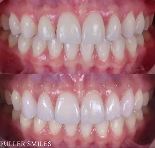
Patient presented to the office with a concern that the teeth were small and that too much gum shows when smiling. A comprehensive exam with x rays, pictures, smile analysis was done.
After understanding the expectations it was decided that the patient needed gum contouring to lift the gums to give bigger teeth to compliment facial anatomy. Temporary veneers were fabricated that were tested by the patient for a week to determine if patient was comfortable with the bite and the way the veneers looked. Finally the hand crafted veneers were fabricated by the lab and meticulously cemented by the doctor to achieve the contour and anatomy that was decided in the beginning of the appointment.
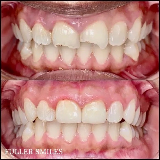
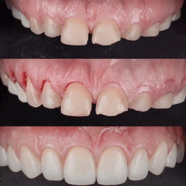
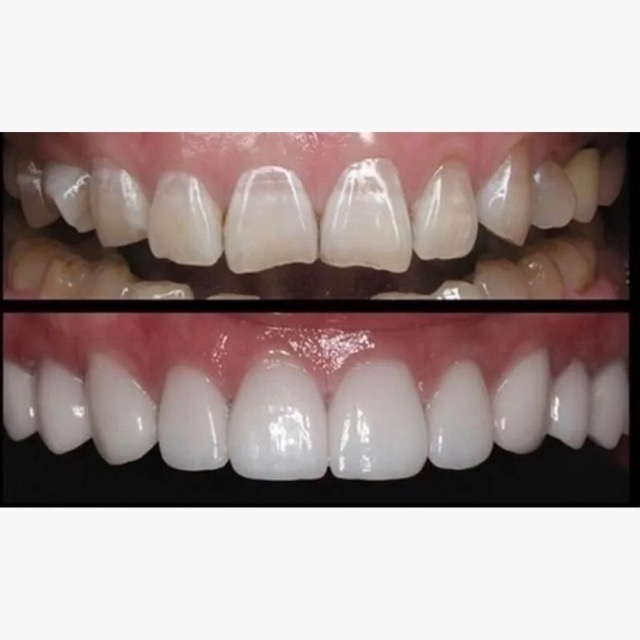
Patient presented to the office with a concern about spaces between the teeth and slight discoloration. A detailed smile analysis with initial pictures, x rays, a scan of the teeth was used to fabricate a mock up smile.
Temporary veneers were fabricated that were given to the patient to test for a week before finally fabricating permanent porcelain veneers to give the teeth a vital, youthful and a natural look.
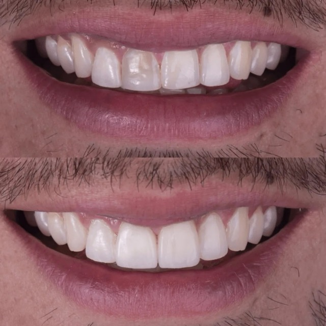
Patient presented to the office with trauma to front two teeth several weeks ago causing her severe pain. After a thorough clinical exam which included endodontic screening and proper x rays led to the diagnosis of necrotic (dead) pulp tissue due to the force of trauma.
Root canals were performed and highly aesthetic Emax crowns were fabricated to meet the patient’s desire to maintain the natural smile and contour as was before the trauma.
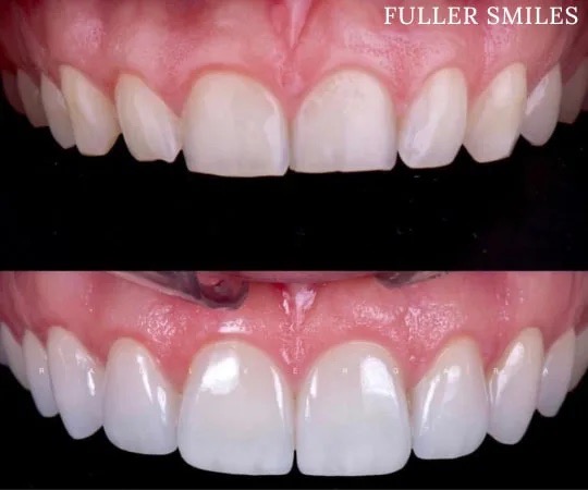
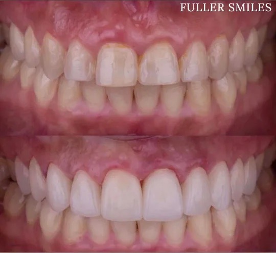
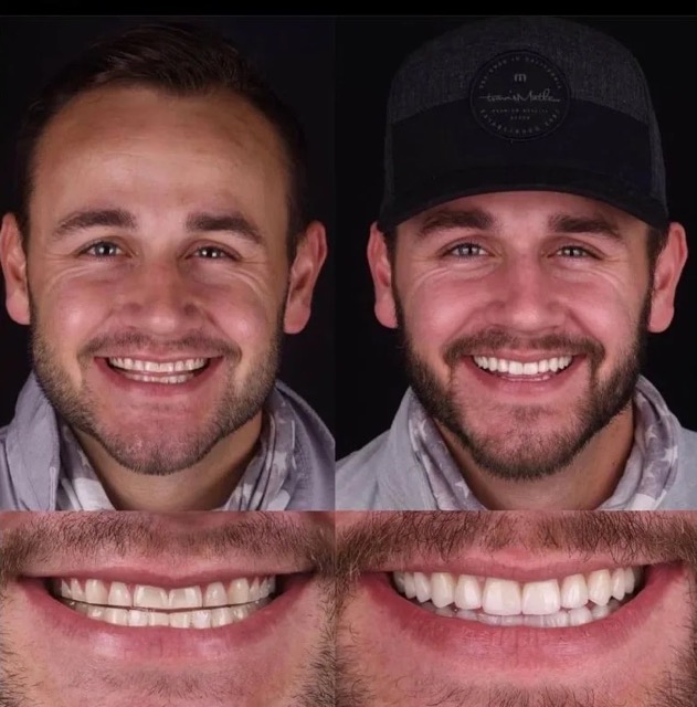
Patient presented to the clinic with the concern about the way smile looked. A thorough aesthetic analysis was done and it was decided that the patient needed bigger teeth and gum contouring to compliment the facial anatomy.
Temporary veneers were then fabricated that were tested by the patient for a week to determine if there were any changes that the patient wanted.Finally naturally looking veneers were hand crafted by the lab that were meticulously cemented to achieve the anatomy and contour that was decided in the beginning of the appointment.
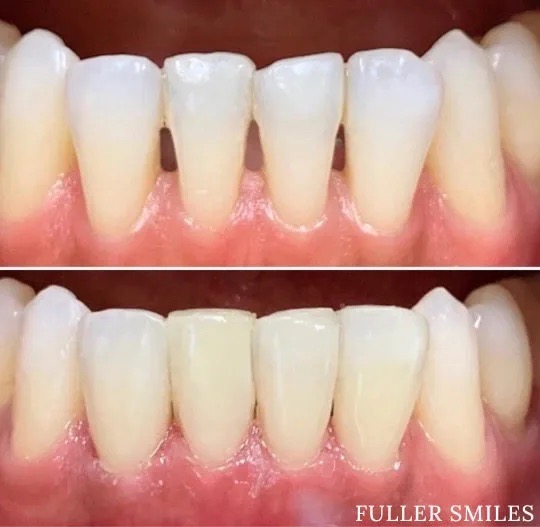
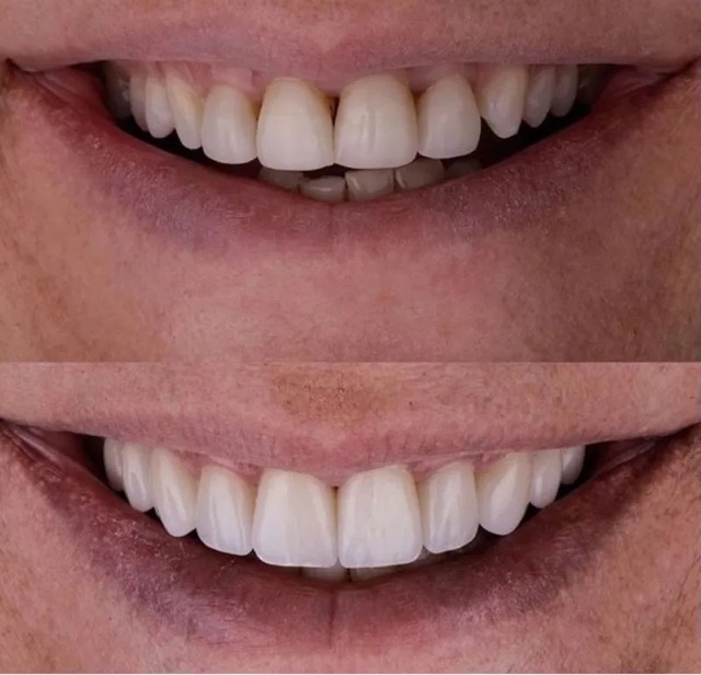
The patient presented to the office with a concern about spaces between lower front teeth. After a detailed conversation with the patient and a thorough clinical exam, the patient decided to get composite bindings done to close the gaps.
Our Dentists used cutting edge technology to get the color and contour right to close the gaps and achieve a highly aesthetic result. Sometimes all it takes is simple tooth bonding.
A thorough clinical exam, endodontic screening and proper x rays led to the diagnosis of irreversible pulpitis(inflamed pulp tissue) due to secondary dental caries with inflamed periodontal ligaments.
Our Dentists used the latest technology and highest quality products to finish this beautiful root canal followed by a post and core to give the tooth additional strength. The tooth was crowned after the root canal to restore function and aesthetics.
Patient presented to the office with a concern about the smile. Patient did not like the way teeth were and was causing severe emotional stress. A detailed smile analysis was performed that included taking multiple pictures, x rays and scanning the teeth to create models to replicate the way patient bites.
A mock up smile was then created on the teeth models to take in patient input. Gum contouring was completed using latest laser technology and temporary veneers were fabricated for the patient to test out for a week. Finally porcelain veneers were fabricated to give the teeth a vital, natural, youthful abs a beautiful appearance.
Patient presented to the office with a concern about front two teeth that had been discolored over time. A detailed clinical exam and smile analysis was performed that included taking multiple pictures, x rays and scanning the teeth to create models to replicate the way patient bites.
One of the discolored teeth had the dead nerve that was treated with root canal and teeth were prepared for veneers. Temporary veneers were fabricated for the patient to test out for a week. Finally porcelain veneers were fabricated to give the teeth a vital, natural and youthful appearance.
Patient presented to the office with a concern that the smile made them look old. A detailed clinical exam, smile analysis and understanding the patient expectations led us to a treatment plan that included 10 minimally prepped veneers to achieve a young and a balanced smile.

A thorough clinical exam, endodontic screening and proper x rays led to the diagnosis of irreversible pulpitis(inflamed pulp tissue) due to secondary dental caries with inflamed periodontal ligaments.
Our Dentists used the latest technology and highest quality products to finish this beautiful root canal followed by a post and core to give the tooth additional strength. The tooth was crowned after the root canal to restore function and aesthetics.
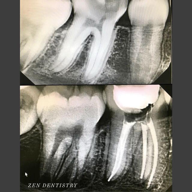
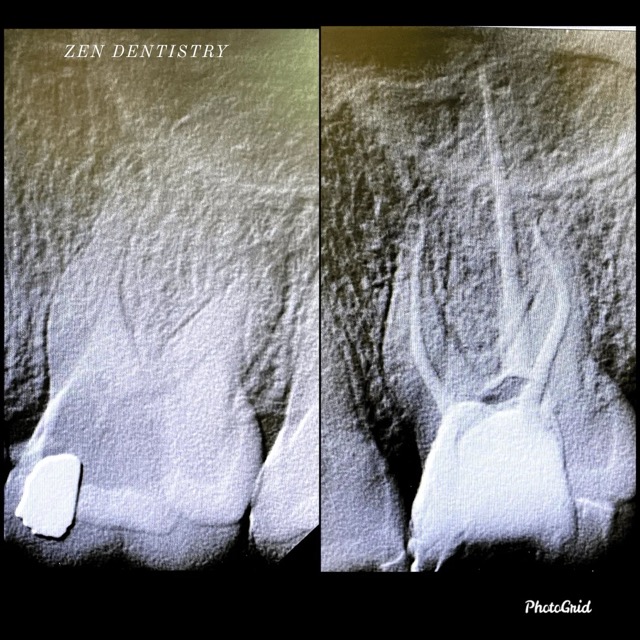
Patient presented to the office with a concern that the tooth has been causing severe constant pain that would become worse at night time and biting. A detailed clinical exams including endodontic screening and x rays let to the diagnosis of pulp necrosis (dead nerve)due to gross dental caries that reached the nerve of the tooth with symptomatic apical periodontitis (inflamed ligaments).
Root Canal treatment was performed that included removing the caries, dead nerve tissue and disinfecting the canals with proper medication to achieve a 3D hermetic seal of the canals to save the tooth.
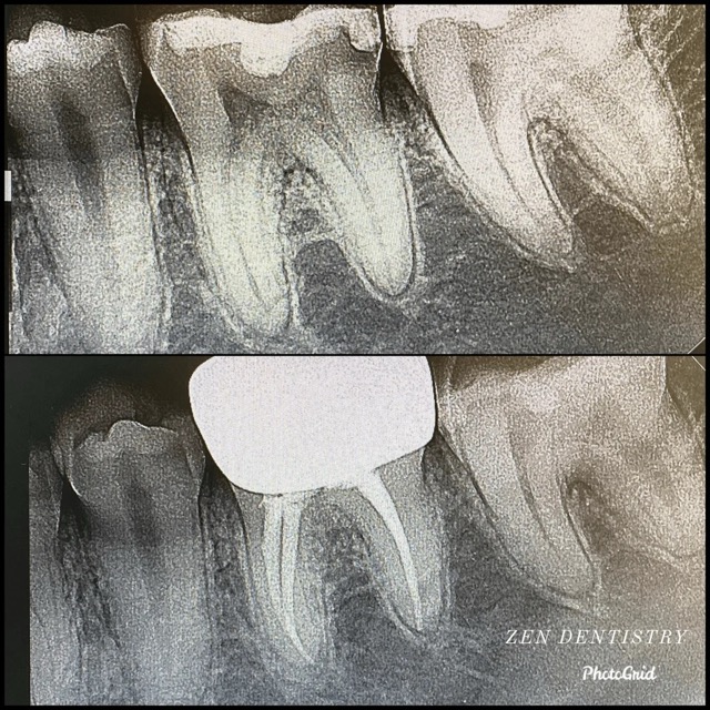
Patient presented to the office with a concern that the tooth has been causing severe constant pain that would become worse at night time and biting. A detailed clinical exams including endodontic screening and x rays let to the diagnosis of irreversible pulpitis (inflammation of the nerve) due to existing filling that was very close to the pulp and symptomatic apical periodontitis (inflamed ligaments).
The tooth was taken out of the bite and pain medication was prescribed to see if the pain could be resolved without having to do a RCT. However tooth responded in the same manner after a week. So it was decided to do a RCT. Root Canal treatment was performed that included removing the caries, dead nerve tissue and disinfecting the canals with proper medication to achieve a 3D hermetic seal of the canals to save the tooth.
Patient presented to the office with a concern that the tooth has been causing severe constant pain that would become worse at night time. A detailed clinical exams including endodontic screening and x rays let to the diagnosis of irreversible pulpitis (infected nerve) due to gross dental caries that reached the nerve of the tooth with asymptomatic apical periodontitis (normal ligaments).
Root Canal treatment was performed that included removing the caries, dead nerve tissue and disinfecting the canals with proper medication to achieve a 3D hermetic seal of the canals to save the tooth.
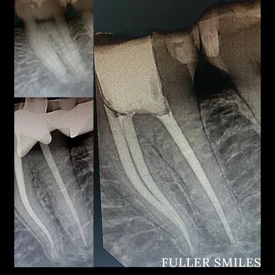
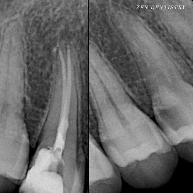
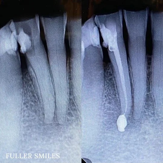
Patient presented with a complaint of severe constant pain that would exaggerate on laying down and biting. Patient wanted to save the tooth. A thorough clinical exam, endodontic screening and proper x rays led to the diagnosis of necrotic (dead) pulp due to dental caries with inflamed periodontal ligaments.
Our Dentists used the latest technology and highest quality products to finish this beautiful root canal. The tooth was crowned after the root canal to restore function and aesthetics.
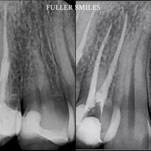
Patient presented with a complaint of severe constant pain that would exaggerate on laying down and biting. A thorough clinical exam, endodontic screening and proper x rays led to the diagnosis of irreversible pulpitis(inflamed pulp tissue) due to secondary dental caries with inflamed periodontal ligaments.
Our Dentists used the latest technology and highest quality products to finish this beautiful root canal followed by a post and core to give the tooth additional strength. The tooth was crowned after the root canal to restore function and aesthetics.
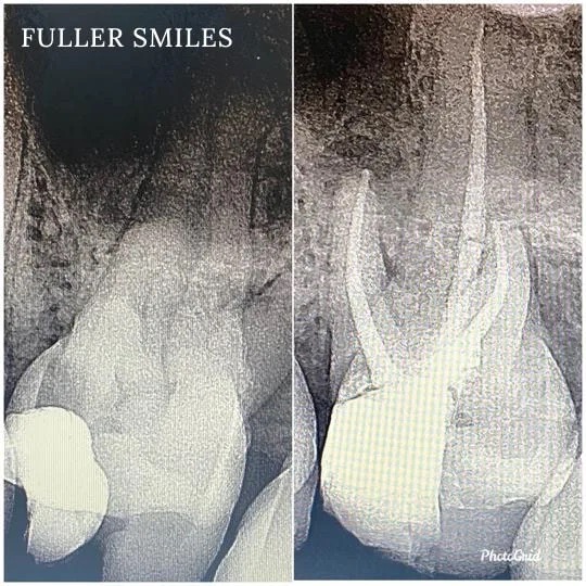
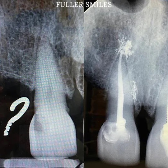
Patient presented with a complaint of severe constant pain that would exaggerate on laying down and biting. A thorough clinical exam, endodontic screening and proper x rays led to the diagnosis of necrotic pulpal tissue(dead pulp tissue) due to secondary dental caries with inflamed periodontal ligaments.
The tooth also had periapical abscess(infection at the bottom of the tooth). Our Dentists used the latest technology and highest quality products to disinfect the root canal system using medication to finish this beautiful root canal. The tooth was crowned after the root canal to restore function and aesthetics.
Patient presented to the office with a concern that the tooth had been hurting constantly since last few days. The pain would become worse at night time and would wake the patient up from sleep.
Detailed clinical exam, endodontic screening and x rays led to the diagnosis of irreversible pulpitis (inflamed nerve) and symptomatic apical periodontitis (inflamed ligaments). A root canal was performed that involved treating the nerve, disinfecting the canals and sealing it with a 3D hermetic seal to save the tooth.
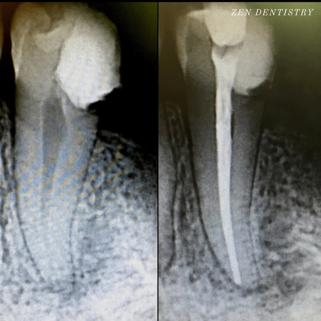
Patient presented with a complaint of severe constant pain that would exaggerate at night time and wake the patient up from sleep.Patient wanted to save the tooth. A thorough clinical exam, endodontic screening and proper x rays led to the diagnosis of irreversible pulpitis due to gross dental caries involving the pulp.
Our Dentists used the latest technology and highest quality products to finish this beautiful root canal. The tooth was crowned after the root canal to restore function and aesthetics.
Patient presented to the office with a concern that the tooth has been causing severe constant pain that would become worse at night time and biting. A detailed clinical exams including endodontic screening and x rays let to the diagnosis of pulp necrosis (dead nerve)due to gross dental caries that reached the nerve of the tooth with symptomatic apical periodontitis (inflamed ligaments).
Root Canal treatment was performed that included removing the caries, dead nerve tissue and disinfecting the canals with proper medication to achieve a 3D hermetic seal of the canals to save the tooth.
A thorough clinical exam, endodontic screening and proper x rays led to the diagnosis of irreversible pulpitis(inflamed pulp tissue) due to secondary dental caries with inflamed periodontal ligaments.
Our Dentists used the latest technology and highest quality products to finish this beautiful root canal followed by a post and core to give the tooth additional strength. The tooth was crowned after the root canal to restore function and aesthetics.
Take a smiling selfie and we’ll show you what Invisalign® treatment can do for you. Sometimes insurances can cover upto $2500 of invisalign treatment. Call our office or follow the link to find out.